Hippocampus
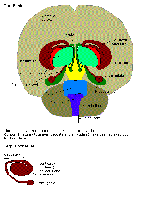
A Hippocampus is a brain structure within a temporal lobe that transfers information into brain memory.
- AKA: Hippocampal Complex.
- Context:
- It can (typically) be composed of a Left Hippocampal Lobe and a Right Hippocampal Lobe.
- It can form episodic representations of the emotional significance and interpretation of events (Phelps, 2004).
- It can be involved in Dreams.
- It can be ascribed to a World Simulator.
- It can support Short-Term Memory, and Long-Term Memory (but not procedural memory).
- …
- Counter-Example(s):
- an Amygdalae.
- a Limbic System.
- See: Cerebral Cortex, Long-Term Potentiation, Temporal Lobe, Navigation, Integrated Brain.
References
2016
- (Wikipedia, 2016) ⇒ http://wikipedia.org/wiki/hippocampus Retrieved:2016-1-29.
- The hippocampus (named after its resemblance to the seahorse, from the Greek ἱππόκαμπος, "seahorse" from ἵππος hippos, "horse" and κάμπος kampos, "sea monster") is a major component of the brains of humans and other vertebrates. Humans and other mammals have two hippocampi, one in each side of the brain. It belongs to the limbic system and plays important roles in the consolidation of information from short-term memory to long-term memory and spatial navigation. The hippocampus is located under the cerebral cortex; [1] and in primates it is located in the medial temporal lobe, underneath the cortical surface. It contains two main interlocking parts: Ammon's horn[2] and the dentate gyrus.
In Alzheimer's disease, the hippocampus is one of the first regions of the brain to suffer damage; memory loss and disorientation are included among the early symptoms. Damage to the hippocampus can also result from oxygen starvation (hypoxia), encephalitis, or medial temporal lobe epilepsy. People with extensive, bilateral hippocampal damage may experience anterograde amnesia — the inability to form and retain new memories.
In rodents, the hippocampus has been studied extensively as part of a brain system responsible for spatial memory and navigation. Many neurons in the rat and mouse hippocampus respond as place cells: that is, they fire bursts of action potentials when the animal passes through a specific part of its environment. Hippocampal place cells interact extensively with head direction cells, whose activity acts as an inertial compass, and conjecturally with grid cells in the neighboring entorhinal cortex.
Since different neuronal cell types are neatly organized into layers in the hippocampus, it has frequently been used as a model system for studying neurophysiology. The form of neural plasticity known as long-term potentiation (LTP) was first discovered to occur in the hippocampus and has often been studied in this structure. LTP is widely believed to be one of the main neural mechanisms by which memory is stored in the brain.
- The hippocampus (named after its resemblance to the seahorse, from the Greek ἱππόκαμπος, "seahorse" from ἵππος hippos, "horse" and κάμπος kampos, "sea monster") is a major component of the brains of humans and other vertebrates. Humans and other mammals have two hippocampi, one in each side of the brain. It belongs to the limbic system and plays important roles in the consolidation of information from short-term memory to long-term memory and spatial navigation. The hippocampus is located under the cerebral cortex; [1] and in primates it is located in the medial temporal lobe, underneath the cortical surface. It contains two main interlocking parts: Ammon's horn[2] and the dentate gyrus.
- ↑ Wright, Anthony. Chapter 5: Limbic System: Hippocampus. Department of Neurobiology and Anatomy, The UT Medical School at Houston
- ↑ Pearce, 2001
2015
- http://psycheducation.org/brain-tours/memory-learning-and-emotion-the-hippocampus/
- QUOTE: So perhaps you would not be surprised to learn that the a portion of the emotion system of the brain (the “limbic system”) is in charge of transferring information into memory. From years of experiments and surgical experience, we now know that the main location for this transfer is a portion of the temporal lobe called the hippocampus.
2007
- (Eichenbaum et al., 2007) ⇒ Howard Eichenbaum, A. R . Yonelinas, and Charan Ranganath. (2007). “The Medial Temporal Lobe and Recognition Memory." Annual review of neuroscience, 30.
- ABSTRACT: The ability to recognize a previously experienced stimulus is supported by two processes: recollection of the stimulus in the context of other information associated with the experience, and a sense of familiarity with the features of the stimulus. Although familiarity and recollection are functionally distinct, there is considerable debate about how these kinds of memory are supported by regions in the medial temporal lobes (MTL). Here, we review evidence for the distinction between recollection and familiarity and then consider the evidence regarding the neural mechanisms of these processes. Evidence from neuropsychological, neuroimaging, and neurophysiological studies of humans, monkeys, and rats indicates that different subregions of the MTL make distinct contributions to recollection and familiarity. The data suggest that the hippocampus is critical for recollection but not familiarity. The parahippocampal cortex also contributes to recollection, possibly via the representation and retrieval of contextual (especially spatial) information, whereas perirhinal cortex contributes to and is necessary for familiarity-based recognition. The findings are consistent with an anatomically guided hypothesis about the functional organization of the MTL and suggest mechanisms by which the anatomical components of the MTL interact to support of the phenomenology of recollection and familiarity.
2004
- (Freeman et al., 2004) ⇒ Hani D. Freeman, Claudio Cantalupo, and William D. Hopkins. (2004). “Asymmetries in the Hippocampus and Amygdala of Chimpanzees (Pan Troglodytes)." Behavioral Neuroscience, 118(6).
- ABSTRACT: Magnetic resonance imaging was used to measure the hippocampal and amygdalar volumes of 60 chimpanzees (Pan troglodytes). An asymmetry quotient (AQ) was then used to calculate the asymmetry for each of the structures. A one-sample t test indicated that there was a population-level right hemisphere asymmetry for the hippocampus. There was no significant population-level asymmetry for the amygdala. An analysis of variance using sex and rearing history as between-group variables showed no significant main effects or interaction effects on the AQ scores; however, males were more strongly lateralized than females. Several of these findings are consistent with results found in the human literature.
- (Phelps, 2004) ⇒ Elizabeth A. Phelps. (2004). “Human Emotion and Memory: Interactions of the Amygdala and Hippocampal Complex." Current opinion in neurobiology 14, no. 2
- ABSTRACT The amygdala and hippocampal complex, two medial temporal lobe structures, are linked to two independent memory systems, each with unique characteristic functions. In emotional situations, these two systems interact in subtle but important ways. Specifically, the amygdala can modulate both the encoding and the storage of hippocampal-dependent memories. The hippocampal complex, by forming episodic representations of the emotional significance and interpretation of events, can influence the amygdala response when emotional stimuli are encountered. Although these are independent memory systems, they act in concert when emotion meets memory.
1997
- (Nadel & Moscovitch, 1997) ⇒ Lynn Nadel, and Morris Moscovitch. (1997). “Memory Consolidation, Retrograde Amnesia and the Hippocampal Complex." Current opinion in neurobiology 7, no. 2
- ABSTRACT: Results from recent studies of retrograde amnesia following damage to the hippocampal complex of human and non-human subjects have shown that retrograde amnesia is extensive and can encompass much of a subject's lifetime; the degree of loss may depend upon the type of memory assessed. These and other findings suggest that the hippocampal formation and related structures are involved in certain forms of memory (e.g. autobiographical episodic and spatial memory) for as long as they exist and contribute to the transformation and stabilization of other forms of memory stored elsewhere in the brain.

